
FIGURE 7.
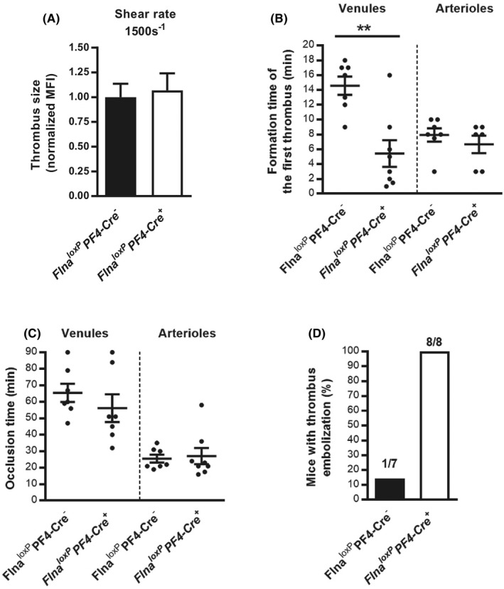
In vivo thrombus instability of Flna loxP PF4‐Cre + mice. The impact of the FlnA mutation on thrombus formation was investigated in vitro. A, A whole‐blood perfusion assay was carried out on collagen matrix (50 μg/ml) at an arterial shear rate (1500 s−1). Thrombus formation was evaluated by fluorescence microscopy and the thrombus size was quantified by measurement of the mean fluorescence intensity (MFI) of thrombi. B‐D, In vivo thrombosis was assessed in exposed mesenteric vessels (venules and arterioles) after FeCl3‐induced injury. Thrombus formation of fluorescently labeled platelets was monitored by intravital videomicroscopy. The graphs represent (B) the time point of the formation of the first thrombus of 30 μm, (C) the occlusion time defined as the stopping blood flow for at least 1 min, and (D) the number of emboli shedding from thrombi in venules throughout the procedure. The graph represents the means ± standard error of the mean of eight Flna loxP PF4‐Cre + and seven Flna loxP PF4‐Cre − mice. Statistical difference between Flna loxP PF4‐Cre − mice and Flna loxP PF4‐Cre + mice was evaluated by the unpaired Student t‐test (**p < .01)