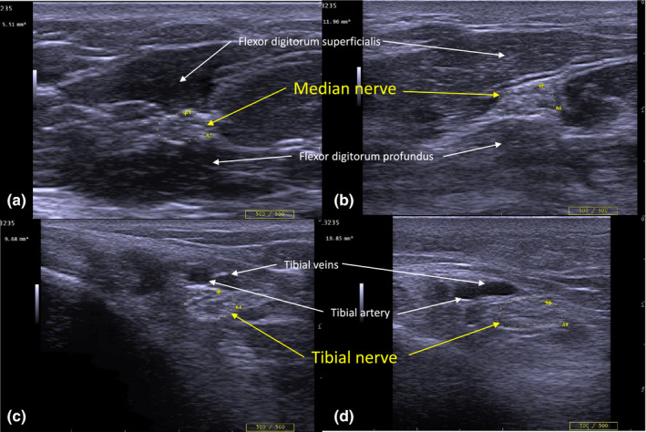
FIGURE 2.

Peripheral nerve ultrasound of median and tibial nerve in diabetes patients with and without neuropathy. Cross‐sectional images of median (a and b) and tibial nerves (c and d) were obtained using peripheral nerve ultrasound. Images a and c are from a patient with type 2 diabetes with a Total Neuropathy Score of 0 and modified Toronto Clinical Neuropathy Score of 0. Images b and d are from a patient with type 2 diabetes with Total Neuropathy Score of 11 and modified Toronto Clinical Neuropathy Score of 16. Nerves on the left‐side panels (a and c) are normal, whereas nerves in the right‐side panels (b and d) are enlarged: median nerve = 11.96 mm2 (normal < 10.01 mm2), tibial nerve = 19.85 mm2 (normal <12.4 mm2). The depth setting for the left‐side image of the tibial nerve is 2 cm, and 3 cm for the right‐side image of the tibial nerve. [Colour figure can be viewed at wileyonlinelibrary.com
