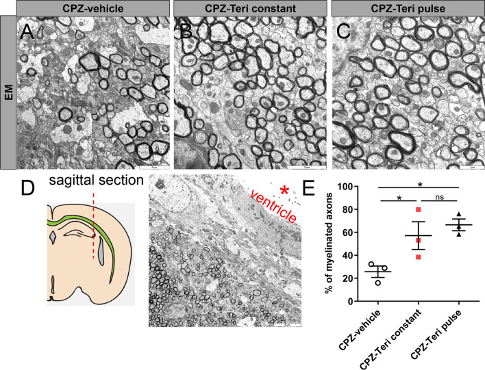
Fig. 4.
A–C Electron micrographs of sagittal corpus callosum (CC) sections from vehicle- and teriflunomide-treated mice showing myelinated axons (n = 3 per group). A, B Scale bars: 2 μM and C scale bar (1 µM). D Region of interest: sagittal section of the CC hitting the axons in cross section. Scale bar 5 µM. E Quantification of myelinated axons in corpus callosum revealed a significant increase upon both stimulation schemes (pulse and constant teriflunomide application). Data are shown as mean values (horizontal lines), while error bars represent standard error of the mean (SEM; vertical lines). Plots also show all individual data points. Significance was assessed using Tukey’s range test following one-way ANOVA (E). Data were considered statistically significant (95% confidence interval) at *p < 0.05, **p < 0.01, ***p < 0.001

