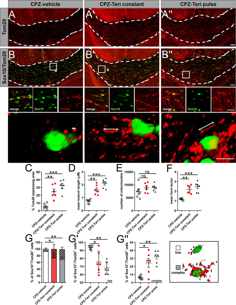
Fig. 6.
Teriflunomide mediated mitochondrial changes in oligodendroglial cells within the corpus callosum. A, B″ Representative images of Sox10 and Tom20 double positive cells in the caudal corpus callosum after 1 week of remyelination. A, B″, C Extent of mitochondrial changes was revealed as the percentage of Tom20-expressing area in the defined region of interest (white borders). Further morphometric quantifications were assessed to determine mitochondrial, D length, E number and F mean form factor (FF). FF = 1 indicates round object, hence low length of mitochondrial networks within cells (form factor 1; low) and increases with elongation, to multiple mitochondria exhibiting elongated tubules (form factor > 1 complex). G–G″ Moreover, the relative percentage of double positive cells (Sox10+ /Tom20+) containing few and less-elongated Tom20+ mitochondria categorized as “low” (blow up; asterix in B) where compared to Sox10-positive cells exhibiting Tom20+ mitochondria with elongated mitochondrial networks categorized as “complex” (blow ups; asterix in B′, B″. G′ Individual data points related to the determination of double positive cells (Sox10 +/Tom20 +) categorized as “low” and (G″) individual data points for double positive cells (Sox10 +/Tom20 +) categorized as “complex”. Number of animals per analysis n = 6. Data are shown as mean values (horizontal lines), while error bars represent standard error of the mean (SEM; vertical lines). Plots also show all individual data points. Significance was assessed using Tukey’s range test following one-way ANOVA (C–G). Data were considered statistically significant (95% confidence interval) at *p < 0.05, **p < 0.01, ***p < 0.001. Scale bars: 50 µm, 20 µm, 10 µm

