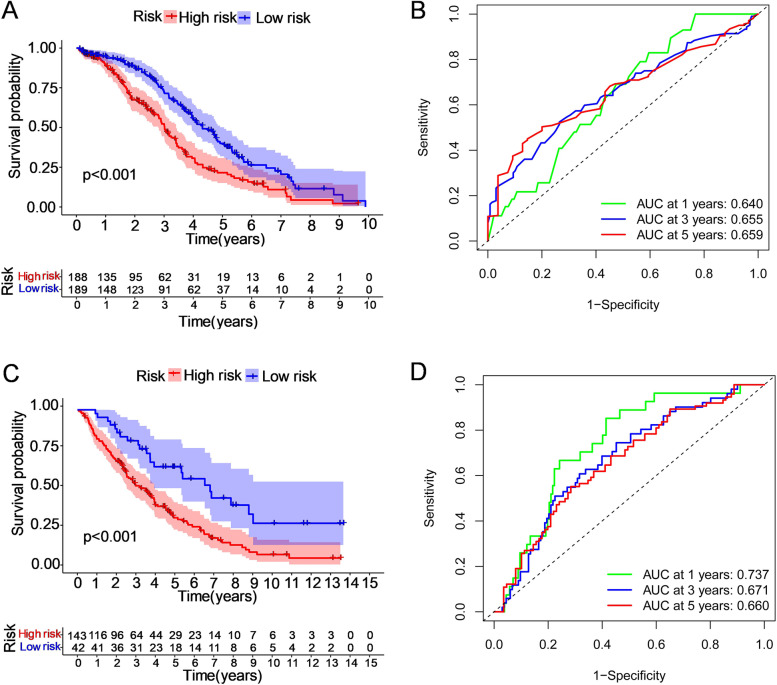
Fig. 4.
The Kaplan‒Meier OS analysis of prognostic models and the ROC curves showing the predictive efficiency of the risk signature. The patients in the two datasets were assigned to the high-risk and low-risk groups (separately represented by red and blue), taking the median risk score as the threshold. A, B In the TCGA discovery set, the survival rate of the high-risk group was lower than that of the low-risk group (P < 0.001). The areas under the curves (AUCs) at 1, 3, and 5 years were 0.640, 0.655, and 0.659, respectively. C, D In the GEO validation cohort, the survival rate was lower for the high-risk group than for the low-risk group (P < 0.001). The areas under the curve (AUCs) at 1, 3, and 5 years were 0.737, 0.671, and 0.660, respectively

