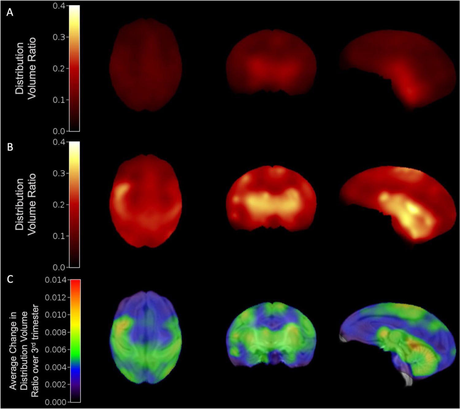
Fig. 3.

Synaptogenesis rates were quantified using average DVR images of the fetal brain. Average (N = 6) DVR images at 120-days (A) and 145-days gestational age (B). C Synaptogenesis rate (SR) image across 6 animals, showing the average change in DVR normalized by the change in gestational age between scans during the third trimester (~ 120- to 145-days gestational age), overlaid on neonate MRI template(21)
