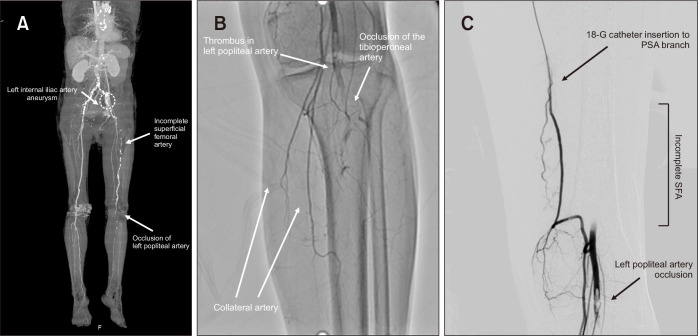
Fig. 1.
(A) Computed tomography angiography showed left superficial femoral artery (SFA) hypoplasia and the thrombus from the left popliteal artery (PA) to the tibioperoneal artery. (B) Conventional angiography revealed a filling defect of left PA. (C) After catheter injection of urokinase, secondary emergent catheter-based thrombolysis was targeted for the left PA thrombus. PSA, persistent sciatic artery.

