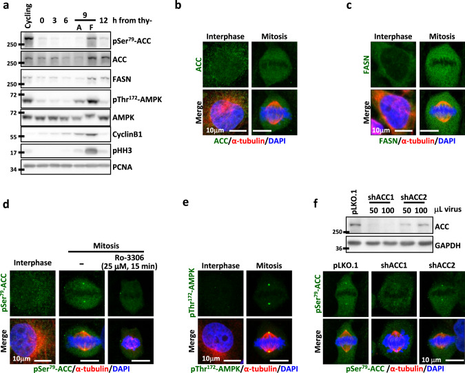
Fig. 1. The expression and cellular distribution of ACC and FASN.
ACC is phosphorylated during mitosis and localizes to the spindle pole (SP). a Level of the indicated proteins at each cell cycle stage. The cell cycle of CGL2 cells was enriched at each stage by double-thymidine block and release. The stages of attached G2 interphase cells (A) and floating mitotic cells (F) collected at 9 h after thymidine release (thy-) were confirmed by the expression of the G2/M markers Cyclin B1 and pHH3. b, c Subcellular localization of ACC (b) and FASN (c) in interphase and mitotic CGL2 cells. Cells were fixed and immunostained for ACC or FASN (green) as described and co-stained for α-tubulin (red) and DAPI (blue). d Subcellular localization of pSer79-ACC in interphase cells, untreated mitotic cells and mitotic cells treated with Ro-3306 as indicated. Cells were stained for pSer79-ACC (green), α-tubulin (red), and DAPI (blue). e Subcellular localization of pThr172-AMPK in interphase and mitotic cells. Cells were stained for pThr172-AMPK (green), α-tubulin (red), and DAPI (blue). f, top: Western blots show the efficiency of ACC depletion by shRNAs. Cells were transduced with shRNA targeting either ACC1 (shACC1) or ACC2 (shACC2) and the lysates were probed with an ACC antibody recognizing both forms. Cells transduced with the empty vector pLKO.1 were used as the control. The volumes of the shRNA-containing virions are indicated. f, bottom: Subcellular localization of pSer79-ACC in mitotic cells after transduction with control vector (pLKO.1), shACC1 or shACC2. Cells were stained for pSer79-ACC (green), α-tubulin (red), and DAPI (blue).

