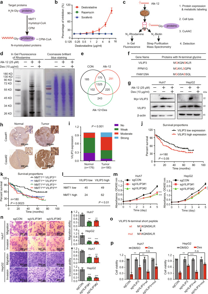
Fig. 4.
Desloratadine inhibits NMT1-mediated myristoylation of the VILIP3 protein. a Schematic diagram showing the approach used to detect NMT1 enzymatic activity. b Detection of NMT1 enzymatic activity in the presence of desloratadine, sorafenib or rapamycin. c Design of the experiment to identify the substrate protein of NMT1. Alk-12-labeled proteins were conjugated to an azido-rhodamine dye for in-gel visualization or with an azido-biotin reagent for click chemistry, followed by purification and analysis by mass spectrometry. d In-gel fluorescence visualization of the myristoylated proteins (left) and the total loaded protein in the NC, Alk-12 and Alk-12+desloratadine groups by Coomassie Brilliant Blue staining (right). e Venn diagram of the mass spectrometry results. f The potential 3 candidate proteins containing a GxxxS sequence. g Western blot showing the level of myristoylated VILIP3 in the presence and absence of desloratadine. h IHC staining showing VILIP3 expression in clinical HCC tissues and adjacent noncancerous tissues. i VILIP3 expression in 180 HCC tumor tissues and 176 matched nontumor tissues. j The VILIP3 expression level was significantly negatively correlated with the overall survival rate of HCC patients (low VILIP3 expression: No and Weak; VILIP3 High expression: Moderate and Strong). k Kaplan–Meier analysis of the correlation between the expression of NMT1/VILIP3 and the overall survival of liver cancer patients. We divided patients into the following four groups, including NMT1low VILIP3low group, NMT1low VILIP3high group, NMT1high VILIP3low group and NMT1 high VILIP3high group. l The expression correlation of NMT1 and VILIP3. m Cell proliferation was evaluated by a CCK-8 assay in the Huh7 and HepG2 cells transduced with sgCON, sgVILIP3#1 or sgVILIP3#2. n The migration and invasion abilities of Huh7 and HepG2 cells expressing sgCON, sgVILIP3#1, or sgVILIP3#2 were evaluated. o Schematic diagram of the mutation site in the VILIP3 protein. p A series of generated Huh7 and HepG2 cell lines were treated with 6 µg/ml desloratadine or DMSO, and cell viability was evaluated using a CCK-8 assay. Bars, SDs; **p < 0.01, ***p < 0.001

