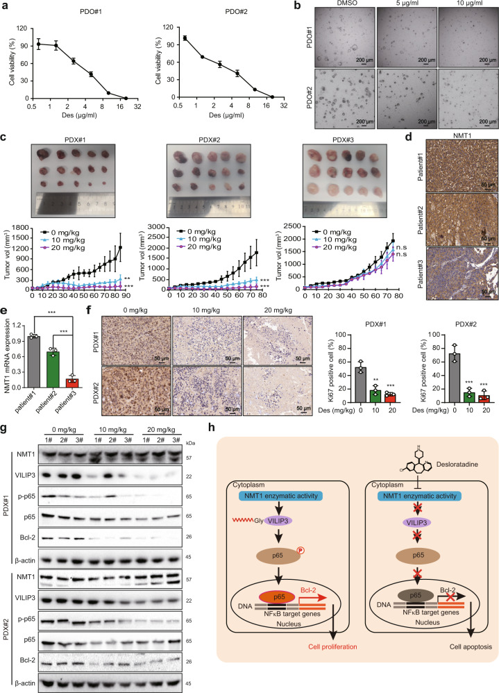
Fig. 6.
Desloratadine suppresses HCC tumorigenesis in PDO and PDX models. a Organoids (PDO#1 and PDO#2) were treated with different concentrations of desloratadine or vehicle (DMSO) for 96 h, and cell viability was evaluated by a CellTiter-Glo Luminescent Cell Viability Assay. b The effect of desloratadine on organoid morphology was monitored. c Representative images showing the tumor growth curves of mice bearing established PDXs. Mice were fed 100 µl of desloratadine suspension (10 mg/kg or 20 mg/kg body weight) once every five days or an equal volume of vehicle (n = 5 per group). d Detection of NMT1 protein expression in HCC tissues from three patients by immunohistochemistry. e Analysis of the mRNA expression level of NMT1 in HCC tissues from three patients by qRT-PCR. f Ki67 staining in tumor tissue sections. g Western blot analysis showing the expression of NMT1, VILIP3, and NFκB signaling pathway-related proteins in the xenograft tumors. h Schematic diagram summarizing how desloratadine suppresses NMT1 enzymatic activity and tumorigenesis. Bars, SDs; *p < 0.05; **p < 0.01, ***p < 0.001, and n.s., no significance

