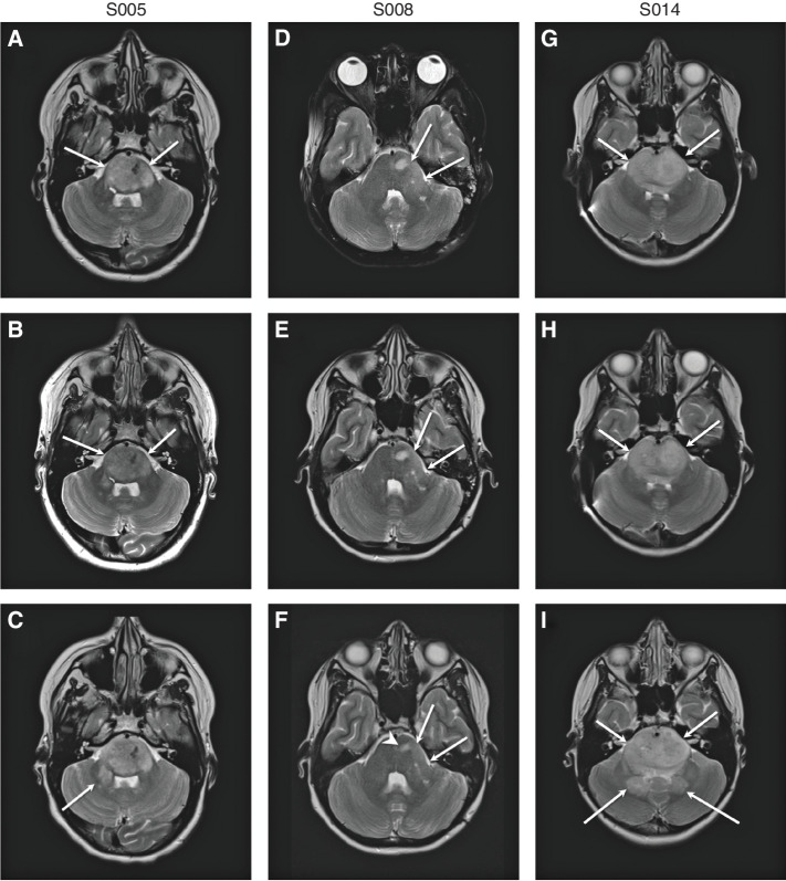
Figure 3.
Neuroimaging after locoregional B7-H3 CAR T-cell infusion. MRI images from S005 (A–C), S008 (D–F), and S014 (G–I). Axial T2-weighted images from all three patients immediately before initial CAR T-cell administration (A, D, and G), following course 2 (i.e., 4 intracranial B7-H3 CAR T-cell infusions; B, E, and H), and prior to any other tumor-directed therapy (C, F, and I). Images correspond to study participants S005 (A–C; imaged at days −2, 53, and 138 relative to first infusion), S008 (D–F; imaged at days −19, 50, and 307 relative to first infusion), and S014 (G–I; imaged at days −1, 45, and 211 relative to first infusion). Patients S005 and S014 experienced slow progression in tumor bulk over time, with increased tumor infiltration in the right brachium pontis (arrow in C) and dentate nuclei (posterior arrows in I). Patient S008 showed mildly decreased tumor bulk (long arrows in F) and decreased conspicuity of T2 hyperintense tumoral nodule (short arrowhead in F).

