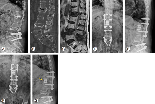
Fig. 3.
A 64-year-old female with an osteoporotic burst fracture at L1. She was treated by vertebral augmentation at another hospital; however, she complained of persistent back pain and gait disturbance. (A) Plain radiograph shows a decreased body height and cement leakage into intervertebral disc space at L1 (yellow arrow). (B) Sagittal computed tomography image display a nonunion and fragment retropulsion into the spinal canal. (C) Sagittal magnetic resonance image (T2-weighted) of lumbar spine displays a compressed spinal cord at L1 (yellow arrow). (D, E) We performed combined anterior and posterior spinal fusion including anterior corpectomy, expandable cage insertion, and multilevel pedicle screw fixations with sublaminar hooks. (F, G) Plain radiographs show the stable status of instruments and complete anterior bony bridge (yellow arrow).

