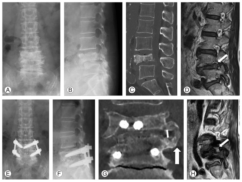
Fig. 4.
Case example for grade 2 collapse. (A, B) Preoperative anteroposterior and lateral radiograph demonstrating grade 2 collapse at L4. (C) Preoperative sagittal computed tomography (CT) image showing inferior endplate collapse at L4. (D) Preoperative sagittal magnetic resonance imaging (MRI) showing foraminal stenosis (white arrow). (E, F) Postoperative radiographs at 24 months after LIF. (G) Postoperative coronal CT image showing bony fusion 24 months postoperatively (white arrow). (H) Postoperative parasagittal MRI shows indirectly decompressed neural foramen (white arrow).

