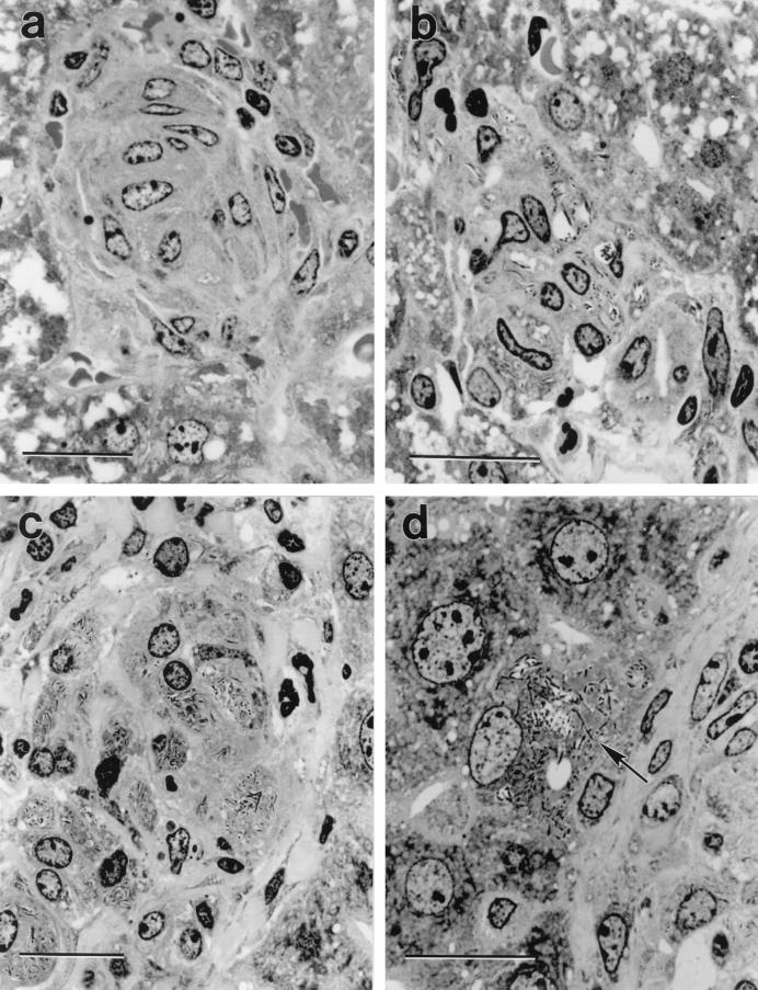
FIG. 1.
Crystal violet-stained, 2-μm-thick plastic section of a liver granuloma. The granuloma in the WT mouse (a) is compact and comprised predominantly of concentrically arranged macrophages, whereas the granuloma in the HC-treated WT mouse (b) is comprised of a loose collection of macrophages containing large numbers of BCG bacilli. The granuloma in the SCID mouse (c) is also comprised predominantly of macrophages that are heavily infected with BCG. The higher power micrograph of the edge of a granuloma in a SCID mouse (d) shows a hepatocyte at the margin of the granuloma (arrow) heavily infected with BCG. Bars represent 20 μm. Magnifications: ×1,060 (a), ×1,280 (b), ×1020 (c), and ×1,280 (d).

