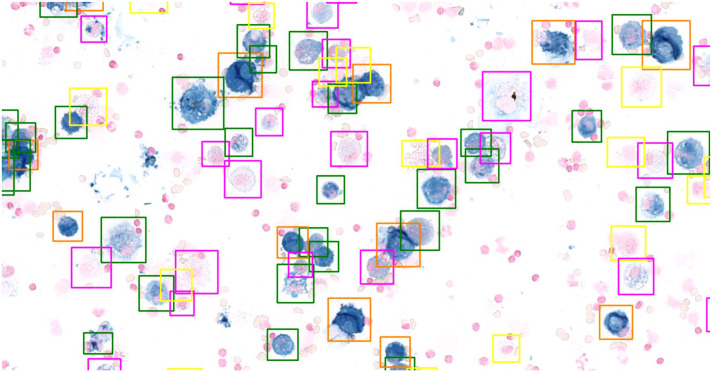
Figure 3.
Cytologic image of a bronchoalveolar lavage fluid stained with Prussian blue. The boxes around the cells represent algorithmic detections of alveolar macrophages with assigned hemosiderin grades according to the scoring system by Doucet and Viel.14 Yellow box, hemosiderin grade 0; pink box, hemosiderin grade 1; green box, hemosiderin grade 2; orange box, hemosiderin grade 3; hemosiderin grade 4 not present in the image.

