FIG. 1.
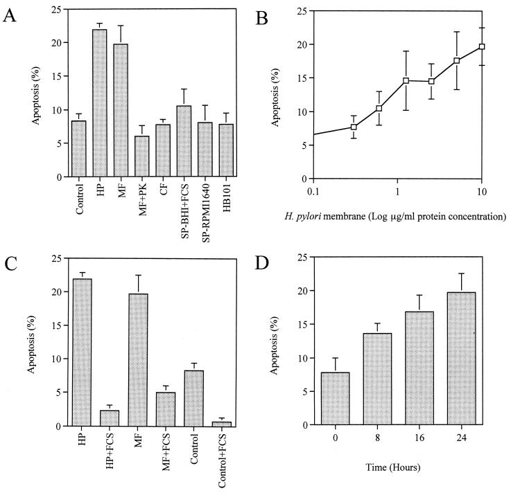
Quantitation of H. pylori-induced apoptosis by fluorescence microscopy. Since there was no significant difference among the strains examined, data from NGY 1281 are shown as representative. Results are expressed as the mean percentages of apoptotic cells per 500 cells enumerated. Bars represent mean values ± SD from three experiments. (A) Induction of apoptosis by live H. pylori (HP), H. pylori membrane fraction (MF), membrane fraction pretreated with proteinase K (MF+PK), H. pylori cytosol (CF), culture supernatant from BHI broth with FCS (SP-BHI+FCS), culture supernatant from RPMI 1640 medium (SP-RPMI 1640), and E. coli HB101 (HB101). (B) Dose-dependent induction of apoptosis by the membrane fraction preparation. (C) Effect of serum on H. pylori-mediated apoptosis. (D) Time-dependent increase in apoptotic activity of the membrane fraction during incubation in serum-free RPMI 1640 medium. Membrane fraction preparations were added at a protein concentration of 10 μg/ml.

