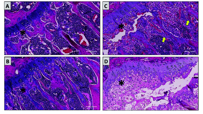
Figure 1.
Tibial physis (asterisks) from a SCID mouse (A), a PXB-mouse supplemented with vitamin C (B), and 2 nonsupplemented (vitamin C deficient) mice (C and D). The primary spongiosa adjacent to the physis of the vitamin C deficient mice had markedly less ossification with microfractures, proliferation of spindle cells along partially mineralized trabeculae (yellow arrows), or complete necrosis of the primary spongiosa (D). The primary spongiosa of supplemented mice (B) euthanized on Day 28 were similar in thickness to the SCID mice (A) with no apparent fractures, spindle cell proliferation, or necrosis.

