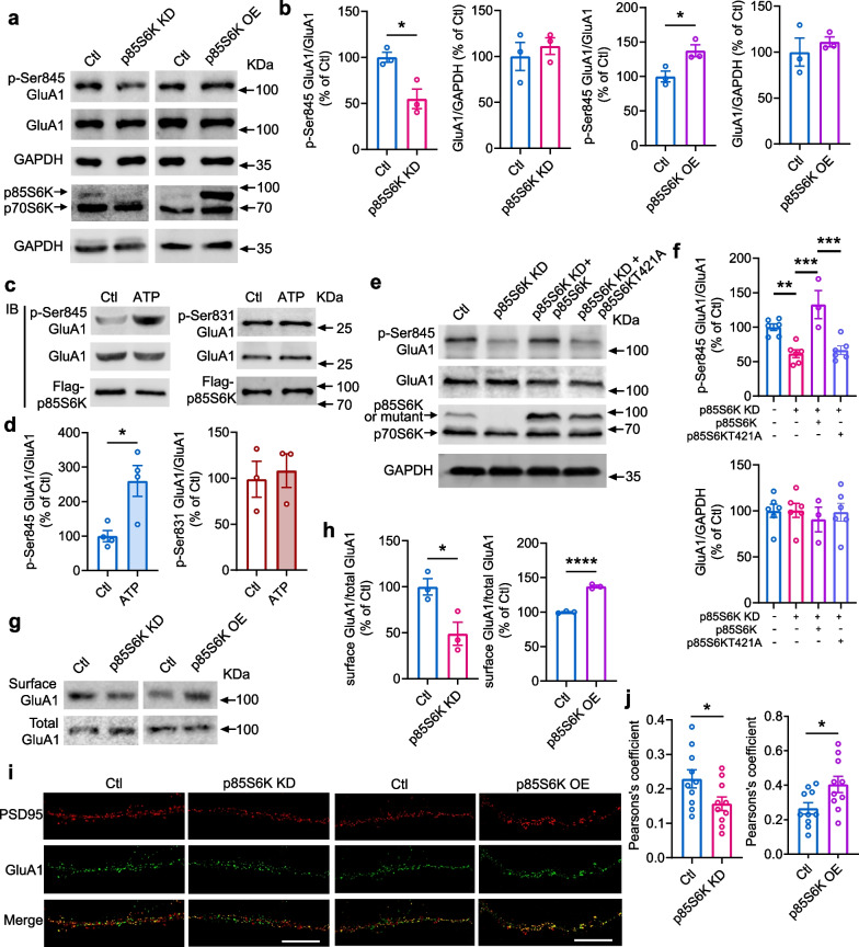
Fig. 3.
p85S6K phosphorylates GluA1 at Ser845 and enhances synaptic GluA1. a, b Knockdown of p85S6K decreased while overexpression of p85S6K increased phosphorylation of GluA1 at Ser845 (p-Ser845 GluA1) in primary hippocampal neurons. Control neurons were infected with lentiviruses expressing control siRNA or empty vectors. n = 3 independent experiments. t = 3.755, df = 4, P = 0.0199; t = 0.6291, df = 4, P = 0.5634; t = 3.234, df = 4, P = 0.0318; t = 0.6827, df = 4, P = 0.5323 (b, from left to right panels). c, d In vitro phosphorylation of GluA1 at Ser845 and Ser831 by p85S6K. n = 4 independent experiments for Ser845 and 3 for Ser831. t = 3.364, df = 6, P = 0.0151 (d, left panel) and Mann–Whitney U = 4, P > 0.9999 (d, right panel). e, f Overexpression of catalytically inactive p85S6KT421A did not rescue the decreased phosphorylation of GluA1 at Ser845 induced by knockdown of p85S6K. n = 3–6 independent experiments. F = 14.52, P < 0.0001. g, h Knockdown of p85S6K decreased while overexpression of p85S6K increased surface GluA1 in primary hippocampal neurons. n = 3 independent experiments. t = 3.312, df = 4, P = 0.0296 (h, left panel) and t = 18.96, df = 4, P < 0.0001 (h, right panel). i, j Colocalization of GluA1 and PSD95 in primary hippocampal neurons overexpressing p85S6K. Scale bar: 10 μm. n = 10 neurons per group. t = 2.168, df = 18, P = 0.0438 (j, left panel) and t = 2.412, df = 18, P = 0.0268 (j, right panel). Data are presented as mean ± SEM. Unpaired t test, two-tailed (b; left panel in d; h; and j), Mann–Whitney test, two-tailed (d, right panel) and ordinary one-way ANOVA followed by Tukey's test (f). *P < 0.05, **P < 0.01, ***P < 0.001, ****P < 0.0001

