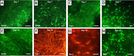
Figure 3.

Major neural cell types contributing to pathology are detectable in cerebellar tissue slices.
Representative fluorescent micrographs of GFAP- (A & B), Tuj-1- (C), and Iba-1-immunoreactive (D) neural cells at 7 days post slice derivation. These cell types, together with MBP-immunoreactive neural cells and potential myelinated fibers, arrows (H), continue to be detectable at 14 days post slice derivation (E–H). Features consistent with neurological injury were occasionally observed in non-lesioned sites: reactive astrogliosis (B; GFAP immunoreactivity) and microglial activation (G; Iba-1 immunoreactivity). FITC: Green; Cy3: red. Scale bars: 50 µm. GFAP: Glial fibrillary acidic protein; Iba-1: ionized calcium-binding adaptor protein-1; MBP: myelin basic protein; Tuj-1: beta tubulin III.
