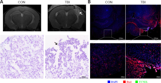
Figure 2.

TBI triggers morphological and cytoarchitectural abnormalities and widespread neuroinflammation at 7 days post-injury.
(A) Top row, representative T2-weighted MRI images of mice from different groups. An elevated T2 signal indicates cerebral edema (arrow). Bottom row, Nissl staining to observe the morphological and cytoarchitectural alterations, which showed TBI-induced neural cell death and gliosis (arrow). (B) Representative images of Iba1 (Cy3, red) immunofluorescence and TUNEL (fluorescein, green) staining. Highly activated microglia and significant levels of apoptosis were observed after TBI. Nuclei (blue) were counterstained with DAPI. Scale bars: 1000 μm (top: lower magnification), 100 μm (bottom: higher magnification). DAPI: 4′,6-Diamidino-2-phenylindole; Iba1: ionized calcium binding adaptor molecule 1; MRI: magnetic resonance imaging; TBI: traumatic brain injury; TUNEL: terminal deoxynucleotidyl transferase dUTP nick end labeling.
