Figure 2.
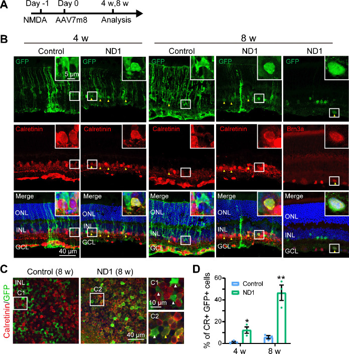
Müller glia are converted into amacrine cells by ND1 in NMDA injured retina.
(A) Illustration of the experimental protocol. (B) Images of retinal sections stained with GFP (green: Alexa Fluor-488) and amacrine cell marker Calretinin (red: Alexa Fluor-647, lane 1–4) or ganglion cell marker Brn3a (red: Alexa Fluor-647, lane 5) at 4 and 8 weeks after virus injection. In the ND1 group, some GFP+ cells co-expressed Calretinin and Brn3a, while there were few such cells in the control group. Yellow arrows point to GFP+ cells that co-expressed neuronal markers. Boxed regions of typical cells are enlarged in the top-right corners of each panel. (C) Images of whole-mount retinas stained with GFP (green) and Calretinin (red: Alexa Fluor-647) in the INL, with white-boxed regions enlarged on the right of the panels. Yellow arrows point to GFP+ cells that co-expressed Calretinin, and white arrows point to GFP+ cells that did not express Calretinin. In the ND1 group, numerous GFP+ cells co-expressed Calretinin, while in the control group few such cells were observed. (D) Percentage of GFP+ cells with Calretinin (CR) among all GFP+ cells at 4 and 8 weeks after virus injection. All data are expressed as mean ± SEM (n = 3 for each group at each time points). *P < 0.05, **P < 0.01 (unpaired Student’s t-test). CR: Calretinin; GCL: ganglion cell layer; GFP: green fluorescent protein; INL: inner nuclei layer; ND1: NeuroD1; ONL: outer nuclei layer.

