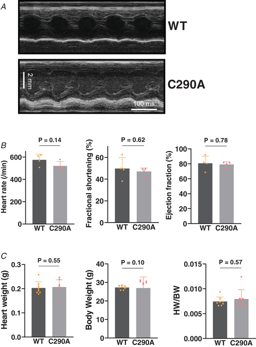
Figure 2. CaMKIIδ‐C290A knock‐in mouse model.

A, representative echocardiogram of the LV in a WT and C290A mouse. B, unaltered heart rate and echocardiographic fractional shortening and ejection fraction in C290A mice. N animal = 4. Unpaired t test. C, normal heart weight, body weight and their ratio in CaMKIIδ C290A mice at baseline. N animal = 8. Mann–Whitney test.
