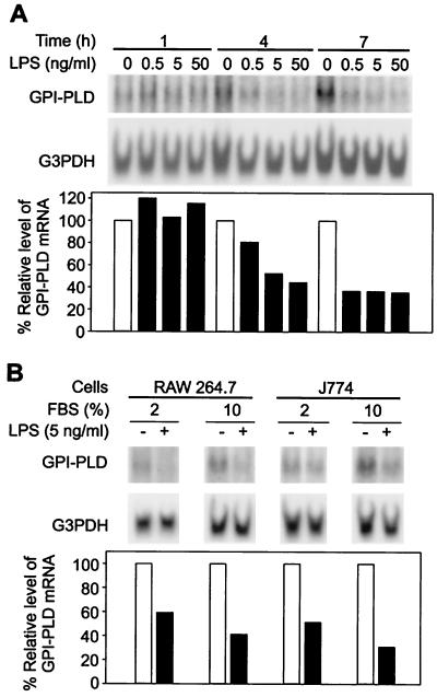
FIG. 1.
Down-regulation of GPI-PLD mRNA by LPS stimulation. (A) Time- and dose-dependent response of GPI-PLD mRNA to LPS stimulation. RAW 264.7 cells were not stimulated (controls) or stimulated with different concentrations of LPS as indicated. Samples were taken at 1, 4, and 7 h for Northern blot analysis using 0.8% RNA denaturing gels. The results are representative of two independent Northern analyses that were hybridized sequentially with 32P-labeled GPI-PLD and G3PDH cDNAs. Intensities of signals, here and in the following figures, were enhanced for presentation purposes; relative levels of GPI-PLD mRNA in stimulated cells in comparison to nonstimulated cells (100%) were calculated as described in Materials and Methods. (B) Reduction of GPI-PLD mRNA by LPS under different serum concentrations. The murine monocyte-macrophage cell lines J774 and RAW 264.7 were stimulated with LPS (5 ng/ml) in medium containing 2 or 10% FBS for 4 h. Total RNA was prepared and analyzed by Northern blotting using 0.8% RNA denaturing gels. Cells without LPS were analyzed in parallel. The results are representative of two independent Northern analyses.

