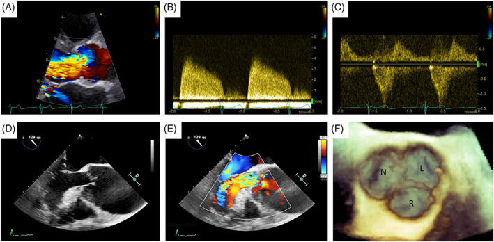
FIGURE 1.

Echocardiographic assessment of aortic regurgitation: (A) TTE colorDoppler focused on left ventricle outflow tract showing a large aortic regurgitant jet, (B) TTE CW Doppler of the regurgitant jet, (C) TTE CW Doppler showing holodiastolic flow reversal in the descending aorta. (D,E) TEE assessment of a patient with severe aortic regurgitation due to a large coaptation defect secondary to aortic root dilatation. (F) 3D TEE assessment of the aortic valve 3D: three dimensional. CW, continuous wave; TTE, transthoracic echocardiography; TEE, transoesophageal echocardiography
