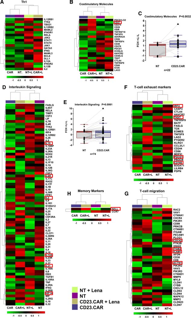
Figure 2. Lenalidomide enhances transcriptional signatures related to T-cell function.
The heat maps and dendrograms of unsupervised hierarchical clustering display differentially expressed genes between NT and CD23.CAR+ T lymphocytes obtained from CLL donor #1, treated in vitro with lenalidomide or left untreated. The gene expression differences among the samples were functionally classified as follows: (A) Th1; (B) costimulatory molecules; (D) interleukin signaling; (F) T-cell exhaustion; (G) T-cell migration; (H) memory markers. Fold changes between lenalidomide (+L) and untreated (-L) sample for costimulatory molecules (C) and interleukin signaling (E) are represented with box and whisker plots.

