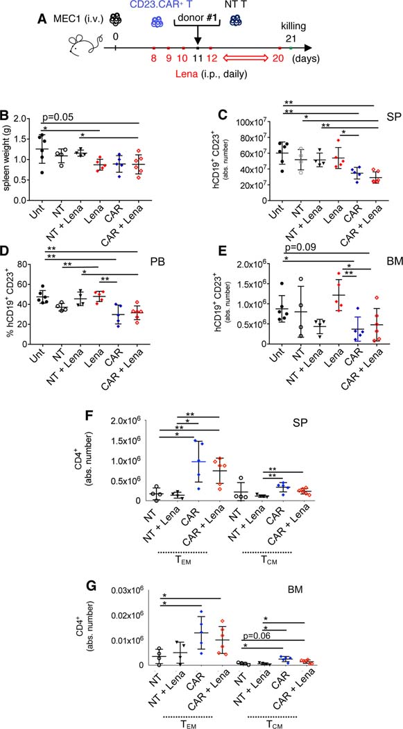
Figure 3. Lymphoid cell compartment characterization in CLL xenotransplanted mice treated with CD23.CAR+ T cells and lenalidomide.
(A-B-C-D-E-F-G) Rag2−/−γc−/− mice transplanted i.v. with MEC1 cells on day 11 of the leukemic challenge were left untreated (Unt, black circles), injected with lenalidomide (Lena) as monotherapy (red rhombi), or adoptively transferred with NT T cells (empty circles), NT T cells with lenalidomide (black triangles), CD23.CAR+ T cells (blue rhombi), CD23.CAR+ T cells with lenalidomide (empty red rhombi). Mice received 0.214 mg/kg of intraperitoneal lenalidomide daily starting at day 8, except for the day of the adoptive transfer. NT and CD23.CAR+ T lymphocytes were obtained from CLL donor #1. At day 23 after the transplantation, mice were evaluated by flow cytometry analysis for the presence of human CD19+ CD23+MEC1 cells in the lymphoid tissues. The graphs show: (B) spleen weight, (C) the mean value (± SD) of the relative contribution of hCD19+ CD23+cells (gated on CD19+ cells) in SP, (D) the mean value (± SD) of the percentage of hCD19+ CD23+cells (gated on CD19+ cells) in PB, (E) the mean value (± SD) of the relative contribution of hCD19+ CD23+cells (gated on CD19+ cells) in BM, (F-G) the mean value (± SD) of the relative contributions of hCD4+ TN, TEM, and TCM in SP and BM.*P < 0.05, **P < 0.01, Student’s t-test.

