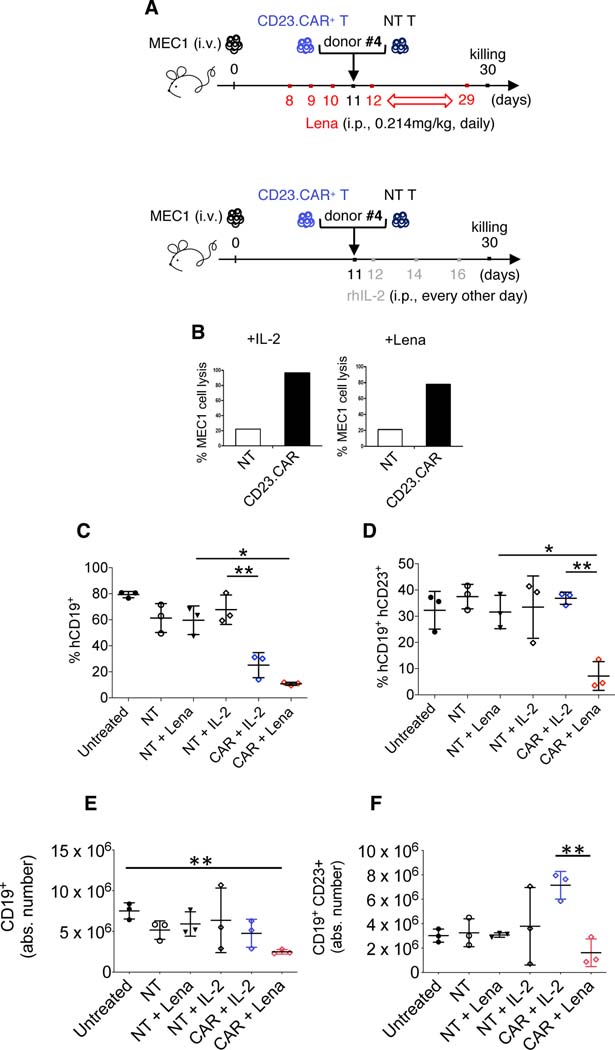
Figure 6. Lenalidomide elicits a tumor-specific cytotoxic activity of CD23.CAR+ T lymphocytes in CLL xenotransplanted mice.
(A-B-C-D) Rag2−/−γc−/− mice who received MEC1 cells intravenously (day 0) were left untreated (black circles) or adoptively transferred with NT T lymphocytes (day 11, white circles), NT T lymphocytes (day 11, black triangles) with daily lenalidomide starting at day 8, NT T lymphocytes (day 11) with rhIL-2 every other day starting at day 12 (six administrations, empty rhombi), CD23.CAR+ T lymphocytes (day 11) with rhIL-2 every other day starting at day 12 (six administrations, blue rhombi), CD23.CAR+ T lymphocytes (day 11) with daily lenalidomide starting at day 8 (red rhombi). NT and CD23.CAR+ T lymphocytes were obtained from CLL donor #4. (B) BM cells were flushed from mice femurs and tibiae; using MACS-microbeads for human CD3 positive selection NT and CD23.CAR+ T cells were isolated via magnetic separation. After 12h of in vitro culture without restimulation, NT (white bar) and CD23.CAR+ (black bar) T cell cytotoxic activity (from IL-2- and lenalidomide-treated mice, respectively) was evaluated against CD23+ MEC1 target, in a 4-hour assay at an E:T ratio of 3:1. The graphs show (C) the mean value (± SD) of the relative contribution of hCD19+ cells in BM, (D) the mean value (± SD) of the relative contribution of hCD19+CD23+ cells in BM, (E) the mean value (± SD) of the absolute number of hCD19+ cells in the BM, and (F) the mean value (± SD) of the absolute number of hCD19+CD23+ cells in the BM. *P < 0.05, **P < 0.01, Student’s t-test.

