Figure1 .
The 5′UTR of cyclin B1 contains an IRES
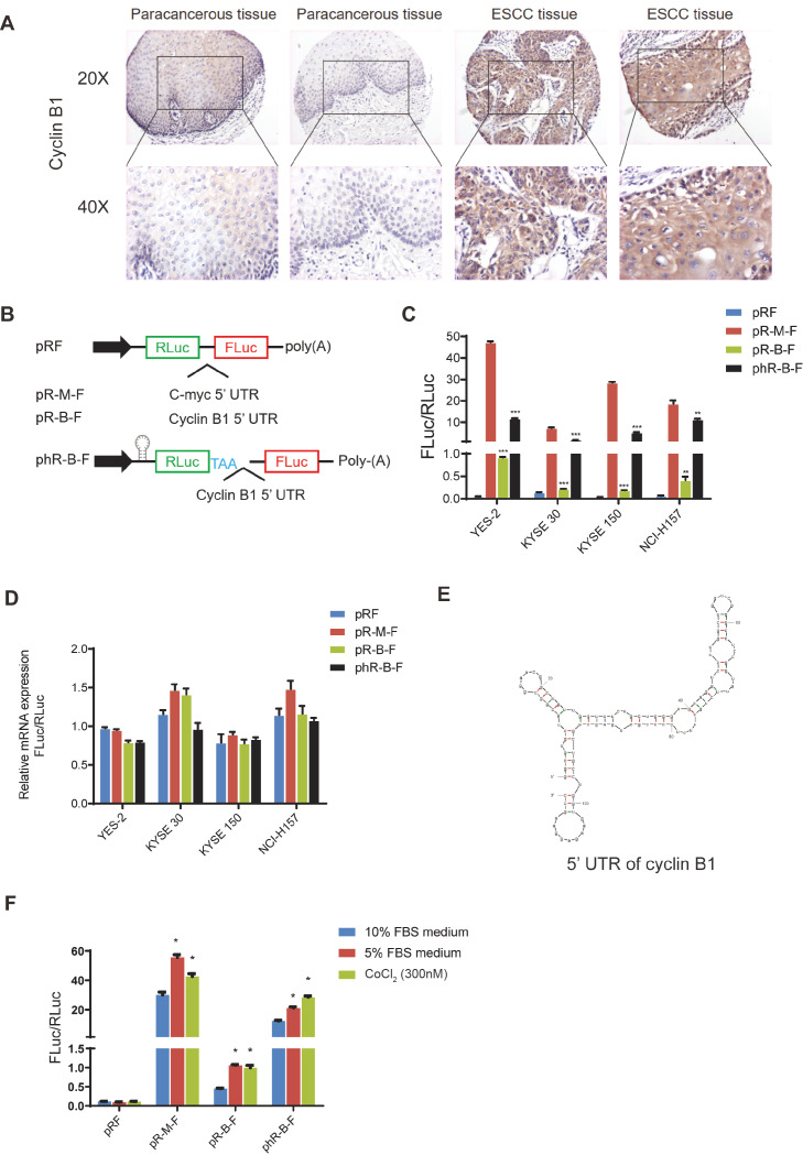
(A) The immunohistochemistry of cyclin B1 in esophageal squamous cell carcinoma (ESCC) and paracancerous tissue. (B) The structure of bicistronic fluorescent reporter plasmid. There are two cistrons in the plasmid. The first cistron is Renilla luciferase (RLuc), and the second cistron is Firefly luciferase (FLuc). Sequences of 5′UTR of cyclin B1 and c-myc are inserted into the plasmid between the two luciferases. A hairpin and an additional stop code are inserted flank the first cistron to prevent the read-through translation. (C) Four cell lines were transfected with the reporter plasmids. Dual-Luciferase® Reporter Assay System was used to measure the activity of the Luciferase. The ratio of FLuc and RLuc indicates the activity of the IRES. Data are presented as the mean±SD of five independent experiments. *P<0.05, **P<0.01, ***P<0.001. (D) The mRNA levels of the two luciferases. RNA was extracted from the four cell lines at 48 h after transfection. Data are presented as the mean±SD of three independent experiments of real-time PCR. (E) The secondary structure of 5’UTR of cyclin B1 mRNA predicted by the RNA Folding Form V2.3. (F) KYSE 30 cells were treated with medium contaning 5% FBS or CoCl2 (300 nM) for 48 h. The ratio of FLuc and RLuc indicates the activity of the IRES. Data are presented as the mean±SD of three independent experiments. *P<0.05.

