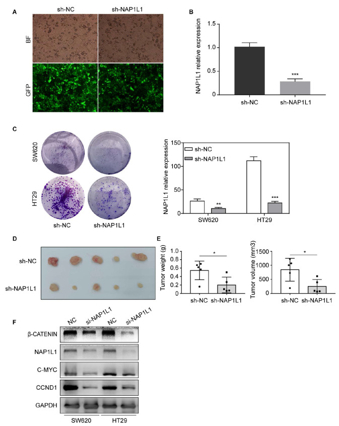
Figure 3 .
Stable knockdown of NAP1L1 suppresses colon cancer growth in vitro and in vivo, and si- NAP1L1inhibits the β-catenin/CCND1 signaling
(A) Images of colon cancer cells transfected with sh-NAP1L1 lentiviral (GFP) were captured after 72 h with a fluorescence microscope. (B) Relative NAP1L1 expression levels were detected by RT-qPCR. *** P<0.001. (C) Colony formation assays were performed after infection with lentiviral particles carrying sh-NAP1L1 precursors or negative control. ** P<0.01, *** P<0.001. (D) Xenograft tumors collected on day 25 after subcutaneous implantation of SW620-NC and SW620-sh- NAP1L1 in nude mice. (E) Tumor volume and weight were measured on Day 25 ( n=5). * P<0.05. (F) β-Catenin, NAP1L1, C-MYC, and CCND1 levels were detected by western blot analysis after transfection with si-NC or si- NAP1L1. GAPDH was used as a loading control.

