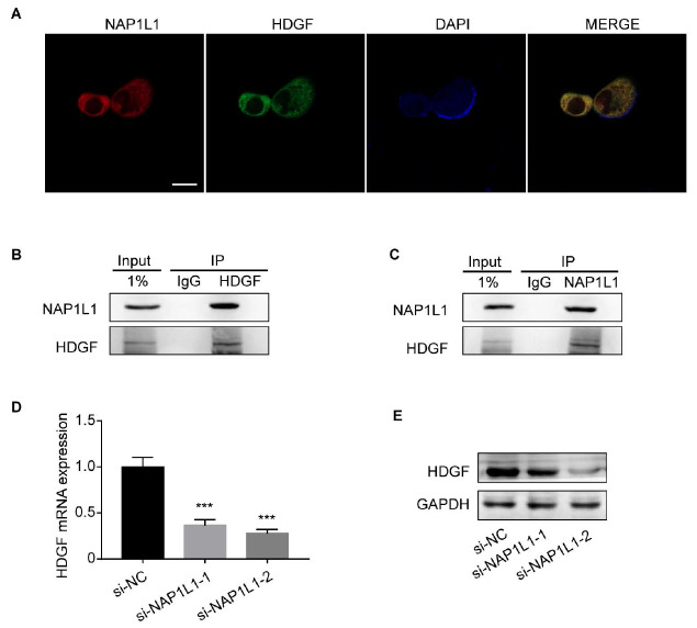
Figure 4 .
Direct interaction of NAP1L1 and HDGF
(A) Immunofluorescence microscopy of NAP1L1, HDGF, and DAPI localization in colon cancer cells. Scale bar: 25 μm. (B,C) Co-IP assays were performed to examine the interaction between NAP1L1 and HDGF. Lysates were immunoprecipitated with anti-HDGF/anti-NAP1L1 antibodies or control IgG, and detected by western blot analysis using anti-NAP1L1 antibodies. (D) HDGF mRNA expression after si-NAP1L1-1/si-NAP1L1-2 transfection, normalized with ADP Ribosylation Factor 5. One-way ANOVA and Dunnett multiple comparison test. *** P<0.001. (E) HDGF protein expression after si-NAP1L1-1/si-NAP1L1-2 transfection. GAPDH was used as a loading control.

