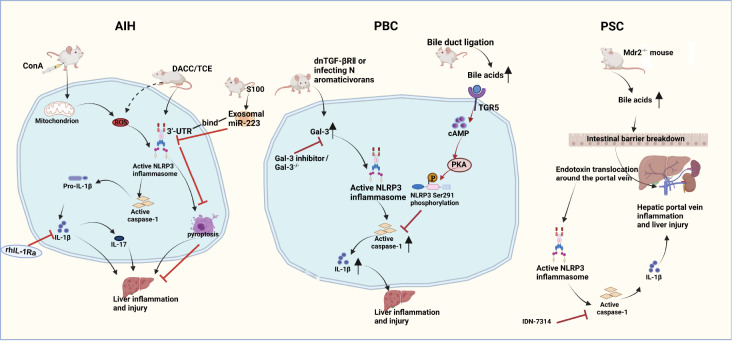
Figure 2 .
NLRP3 inflammasome activation in the disease processes of AIH, PBC and PSC
The NLRP3 inflammasome is activated and IL-1β is upregulated in ConA- or TCE-induced AIH model animals, which ultimately leads to the development of liver inflammation and injury that can be blocked by the IL-1 inhibitor rhIL-1Ra. Overexpressed miR-233 in exosomes from bone marrow-derived stem cells inhibits NLRP3 activation by binding to its 3′UTR, leading to NLRP3 mRNA degradation and thus suppression of liver inflammation and cell death, mediating a form of hepatoprotection. In a spontaneous PBC dnTGF-βRII mouse model and an induced PBC mouse model obtained by infection with Novosphingobium aromaticivorans, Gal3 promotes increased inflammation by enhancing the activation of the NLRP3 inflammasome and IL-1β production, inducing liver inflammation and injury that can be stopped by Gal3 inhibitors. Bile duct ligation triggers elevation in BAs, activating the TGR5-cAMP-PKA pathway and inducing NLRP3 Ser291 phosphorylation, inhibiting caspase-1 activation and relieving liver damage. In a mouse Mdr2-knockout ( Mdr2 −/−) model that duplicates the human PSC, BAs augment intestinal barrier disruption. Impaired barrier function leads to bacterial endotoxin migration to the hepatic periportal vein and activation of the NLRP3 inflammasome, ultimately leading to liver damage. Treatment with the pancaspase inhibitor IDN-7314 ameliorates liver injury in Mdr2 −/− mice.

