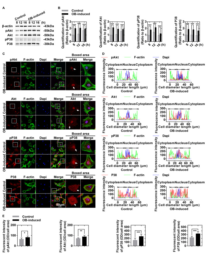
Figure4 .
Osteoblasts regulate glucose-derived ATP production through the Akt and P38 signaling pathways in chondrocytes
(A) Western blots showing the upregulation of pAkt, total Akt, pP38, and P38 in chondrocytes induced by osteoblasts. Cell lysates were collected at 8, 12, and 16 h after co-culture. The gels are representative of three independent experiments (n=3). (B) Quantification was performed to analyze changes in pAkt, total Akt, pP38, and total P38 in (A). (C) Immunofluorescence staining of pAkt, total Akt, pP38, and P38 (red) in chondrocytes after 8 h of co-culture with osteoblasts. The cytoskeletons were stained with FITC-phalloidin (green), and the nuclei were stained with DAPI (blue). The images observed by CLSM were from three independent replicates (n=3). (D) Image-Pro Plus 6.0 was used to determine the linear fluorescence intensity and explore the distributions of pAkt, total Akt, pP38, and P38 in (C). Data analysis was performed on at least 10 cells per group. (E) Fluorescence quantification was performed to show the changes in pAkt, total Akt, pP38, and P38 in (C). The results were from three independent experiments (n=3). The data in B and E are presented as the mean±SD. A significant difference was observed in control chondrocytes. *P<0.05, **P<0.025, and ***P<0.01. OB, osteoblasts.

