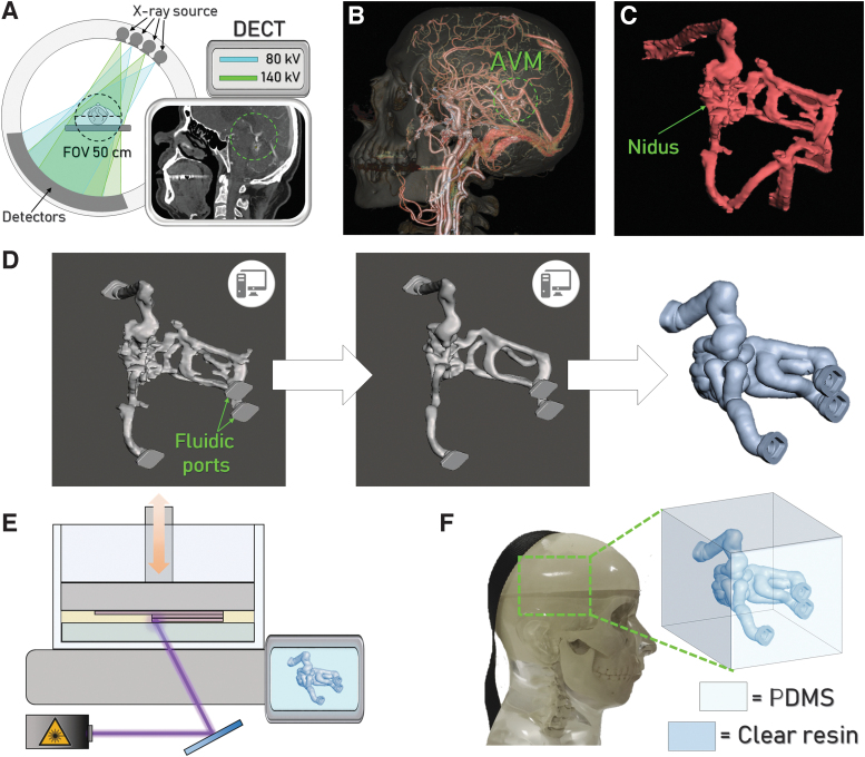
FIG. 1.
Workflow adopted for the vascular phantom realization. (A) Schematic illustration of the DECT scanner employed for CT imaging and a representative image acquired. The green circle highlights the presence of the AVM. (B) 3D rendering elaborated from CT images, used to identify and isolate the AVM (green circle) from the surrounding anatomical structures. (C) 3D solid model of the AVM and (D) 3D model elaboration using MeshMixer® to obtain the hollow AVM. (E) Schematization of the printing process to produce the physical model of the vascular AVM. (F) Representation of the complete phantom constituted by the AVM embedded into the silicone-based cube, fitting the commercial anthropomorphic phantom. 3D, three-dimensional; AVM, arteriovenous malformation; CT, computed tomography; DECT, double energy computed tomography.

