FIG. 6.
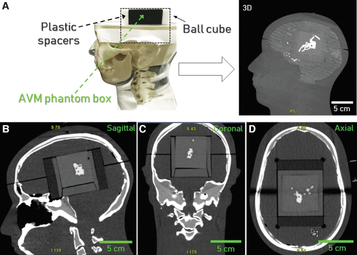
(A) Experimental setup for the validation of the AVM phantom embedded in PDMS filled with the contrast agent and fitted into the anthropomorphic phantom. (B) Sagittal, (C) coronal, and (D) axial CT scans of the phantom. AVM, arteriovenous malformation; CT, computed tomography; PDMS, polydimethylsiloxane.

