Figure 3.

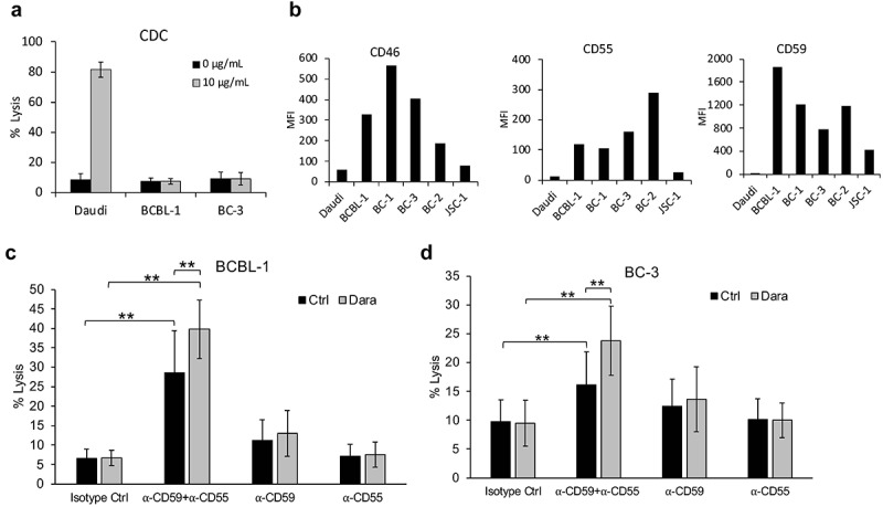
Lack of Dara-mediated complement-dependent cytotoxicity (CDC) of PEL cell lines. (a) CDC in the presence of 20% pooled normal human serum was performed on Daudi, a BL cell line, and BCBL-1 and BC-3 PEL lines, in the presence or absence of 10 µg/mL Dara. Cells were stained with propidium iodide (PI) and % lysis was determined based on PI-positive cells using flow cytometry after 2 h. (b) Levels of complement-inhibitory proteins CD46, CD55, and CD59 on the surface of Daudi or various PEL cell lines were measured by flow cytometry after staining the cells with PerCP-Cy5.5-conjugated anti-CD46 or anti-CD55 antibodies or FITC-conjugated anti-CD59 antibody. Data are expressed as median fluorescence intensity (MFI). (c and d) BCBL-1 (c) and BC-3 (d) cells were incubated with 10 µg/mL isotype control or neutralizing antibodies against CD59, CD55, or both prior to performing CDC assays with or without 10 µg/mL Dara as described in (a). Data are presented as % lysis as determined by PI-positive cells using flow cytometry. Error bars represent standard deviations from at least 3 independent experiments. Statistically significant differences (*P ≤ .05, **P ≤ .01) by 2-tailed t-test are indicated.
