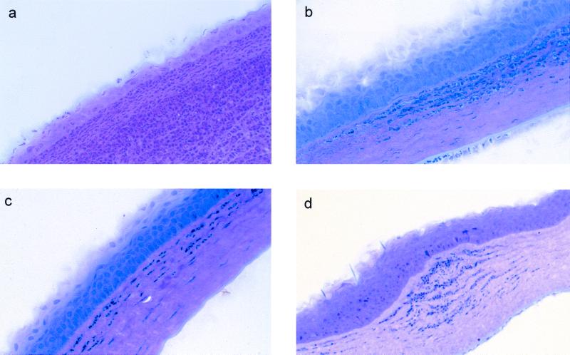
FIG. 2.
Histological examination at 24 h postchallenge of nonimmunized and immunized rat corneas inoculated with cytotoxic P. aeruginosa strain 6206. Panels: a, corneas of nonimmunized rats showing massive PMN infiltration streaming through the limbus and conjunctiva into the mid-periphery of the corneal stroma, with fewer PMNs in the central cornea, bacteria that could be seen throughout the corneal stroma, and thinned epithelium in the periphery and mid-periphery of the cornea; b, corneas of orally immunized rats (50%) showing infiltrates in the periphery of the cornea, with fewer PMNs in the central cornea and healed epithelium at the original scratch site; c, corneas of nasally immunized rats (25%) showing diffuse infiltrates all over the corneal stroma, with the scratch site being healed completely and with no epithelial defect seen at 24 h post challenge; d, IPP-immunized rat corneas (25%) showing few patches of focal infiltrates in the mid-periphery of the corneal stroma and diffuse infiltrates in the periphery of the cornea, with bacteria that could be seen near focal infiltrates in the corneal stroma.

