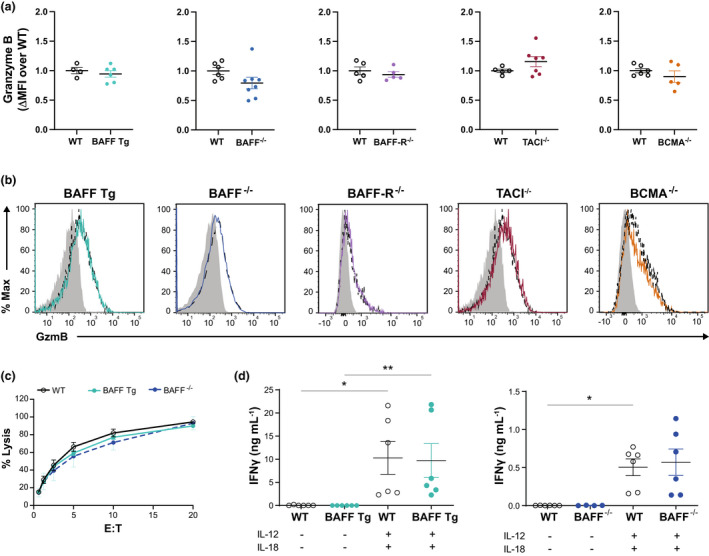
Figure 3.

Analysis of NK cell function ex vivo. (a) ΔMFI of GzmB = geometric MFI of the CD3−NK1.1+ population – geometric MFI of the CD3−NK1.1− population. As the data were pooled from two or three independent experiments, the ΔMFI of each mouse was normalized as the fold‐change over the average ΔMFI of the WT mice in each experiment. (b) Histograms documenting the expression of GzmB in the CD3−NK1.1+ population from GM mice (solid line) compared with WT controls (dashed line). The shaded histograms document GzmB expression in the negative control (CD3−NK1.1−) cell population. (c) 51Cr release assay performed on resting NK cells isolated from the spleens of WT, BAFF transgenic (Tg) and BAFF−/− mice at various E:T ratios. (d) NK cells (CD3−NK1.1+) were isolated at over 95% purity and cultured at 106 cells mL−1 with or without IL‐12 (1 ng mL−1) and IL‐18 (5 ng mL−1). Supernatants were collected at t = 6 h and analyzed using a LEGENDplex cytometric bead array to detect IFNγ. The data (mean ± standard error of the mean) presented in a (n = 5–14 mice) and d (n = 4–6 mice) are pooled from two or three independent experiments. The data presented in b (n = 5–14 mice) and c (n = 4 mice) are representative of two or three independent experiments. *P ≤ 0.05, **P ≤ 0.01. BAFF, B‐cell–activating factor; BAFF‐R, B‐cell–activating factor receptor; BCMA, B‐cell maturation antigen; E:T, effector‐to‐target ratio; GM, genetically modified; GzmB, granzyme B; IFN, interferon; IL, interleukin; MFI, mean fluorescence intensity; NK, natural killer (cells); TACI, transmembrane activator and calcium modulator and cyclophilin ligand interactor; WT, wild type.
