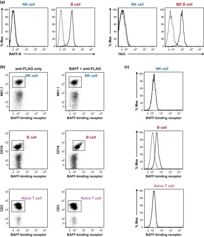
Figure 5.

Flow cytometry analysis of receptor expression on resting NK cells from WT mice. (a) Surface expression of BAFF‐R and TACI on splenic NK cells. Left: solid (WT) and dotted (BAFF‐R−/−) lines represent BAFF‐R expression on NK cells from WT and BAFF‐R−/− mice, respectively, with B cells as positive controls. Right: solid and dotted lines represent the results of staining NK cells from WT mice with an anti‐TACI antibody and fluorescence minus one control, respectively. Splenic MZ B cells (CD19+IgMhighCD21/35highCD23−CD93−) were used as positive controls. The data shown are representative of three experiments that yielded similar results. (b) Splenocytes from WT mice were incubated either with or without FLAG‐tagged mouse recombinant BAFF followed by anti‐FLAG M2 FITC secondary antibody. CD19+ B cells and naïve T cells (CD19−CD3+CD44lowCD62Lhi) served as positive and negative controls, respectively. NK cells were identified as CD19−CD3−NK1.1+. (c) Histograms document the binding of recombinant BAFF to NK, B and naïve T cells. Dotted and solid lines denote cells detected only with anti‐FLAG and cells detected with recombinant BAFF together with anti‐FLAG, respectively. The data shown are representative of two independent experiments with 2 mice per group (n = 4 mice). BAFF, B‐cell–activating factor; BAFF‐R, B‐cell–activating factor receptor; FITC, fluorescein isothiocyanate; MZ, marginal zone; NK, natural killer (cells); TACI, transmembrane activator and calcium modulator and cyclophilin ligand interactor; WT, wild type.
