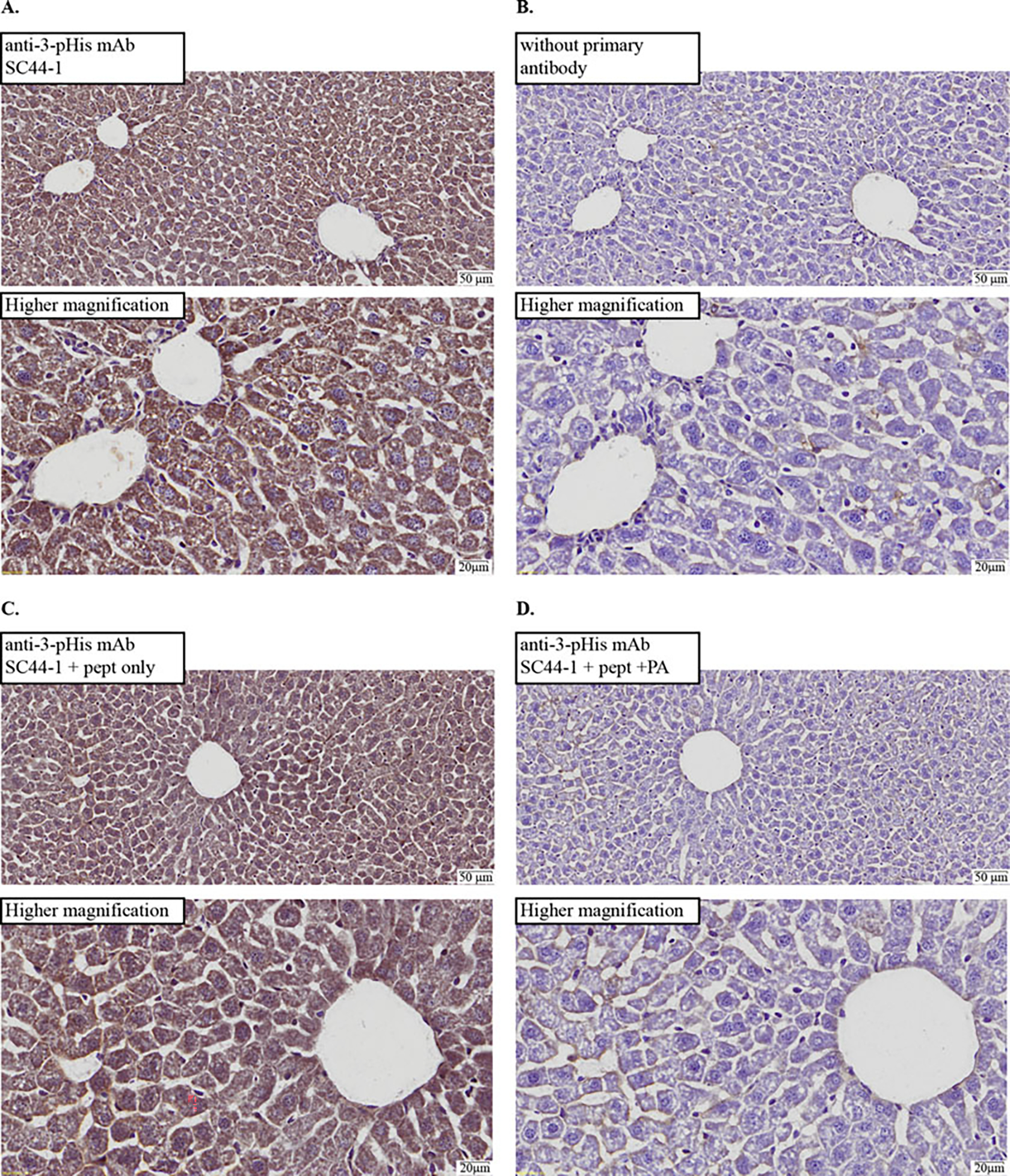
Fig. 2.

Chromogenic detection of SC44-1 anti-3-phosphohistidine mAb signals in cryosections from formaldehyde-fixed, cryoprotected mouse liver. IHC images of slides for serial cryosections of C57BL/6 normal mouse liver. To best compare controls to experimental conditions, two separate slides were processed and analyzed: one was mounted with both (a) and (b) cryosections and another mounted with both (c) and (d) cryosections. Low magnification (top) and higher magnification (bottom) images with a scale bar are shown for each. Histidine phosphorylated peptides or controls were used in primary antibody incubations to probe phosphorylation-dependence of the IHC signal: (a) antibody (SC44-1) incubated without peptide (pept); (b) blocking buffer alone (no antibody) used in primary incubation; (c) antibody incubated with pept, untreated with phosphoramidate (PA); (d) antibody incubated with pept treated with PA (i.e., phosphorylated). Results support the phosphorylation dependence of IHC signal
