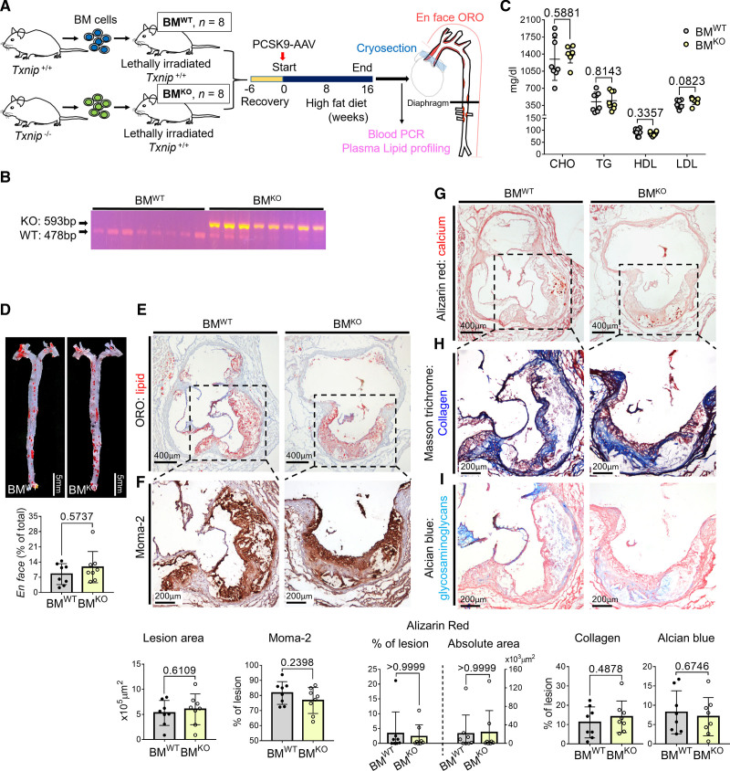
Figure 3.
Hematopoietic deficiency of TXNIP (thioredoxin-interacting protein) does not affect the lesion phenotype. A, Schematic illustration of the experiment. Bone marrow (BM) cells of wild type (WT) or Txnip knockout mice (Txnip KO) were transplanted into WT mice. After 6 weeks of recovery, the mice were injected with PCSK9-AAV and fed HFD for 16 weeks. n=8/8 for bone marrow cells of wild type mice (BMWT)/ bone marrow cells of Txnip KO mice (BMKO). B, Blood PCR result indicating the successful transfusion of the WT and Txnip KO BM cells. C, The plasma concentrations of total cholesterol (CHO), triglyceride (TG), high-density lipoprotein (HDL), and low-density lipoprotein (LDL). D, Comparison of the Oil Red O (ORO)-positive areas between BMWT and BMKO on en-face aorta. E–I, Characterization of atherosclerotic lesions using the 7 μm of perpendicular serial sections (total 70–80 sections) prepared from the aortic sinus. Representative serial sections and quantification results of Oil Red O (ORO) staining (E, for lesion size), Moma-2 immunostaining (F, for monocyte/macrophage), Alizarin red staining (G, for calcium), Masson trichrome staining (H, for collagen), Alcian blue (I, for glycosaminoglycan ECM). The applied statistical tests and results are summarized in the Table S11. The error bars denote standard deviation. The exact P values are specified.

