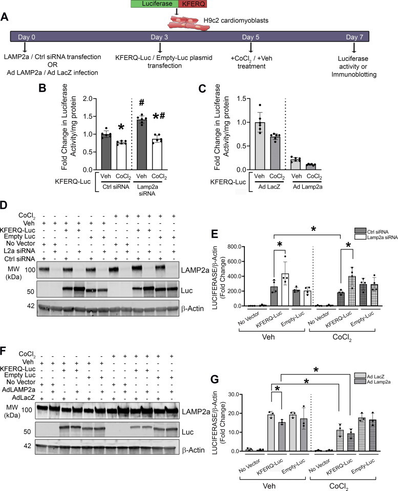
Figure 5.
Changes in chaperone-mediated autophagy (CMA) by luciferase reporter assay with altered lysosome associated membrane protein receptor 2a (LAMP2a) and hypoxic stress. H9c2 cells with Lamp2a knockdown or overexpression (and corresponding controls) were transfected with either a KFERQ-luciferase (KFERQ-Luc) CMA reporter plasmid or control Empty-luciferase (Empty-Luc) plasmid for 72 h. An additional negative control group of cells, not transfected with any plasmid, was added to each experiment. The cells were then treated with CoCl2 or vehicle (Veh) for an additional 48 h. A: a schematic timeline for KFERQ-Luc or Empty-Luc transfection and Veh or CoCl2 treatment. B: bar graph shows fold change in luciferase activity observed in the Lamp2a silenced H9c2 cells with CoCl2 or Veh. C: bar graph shows the fold change in luminescence from Lamp2a-overexpressing H9c2 cells with CoCl2 or Veh, transfected with KFERQ-Luc (n = 6 independent samples/group). D: representative immunoblots of LAMP2a, luciferase (Luc), and β-actin protein levels are shown from Lamp2a silenced H9c2 cells and (E) represents the quantification of Luc protein levels normalized to β-Actin levels (n = 4 samples/group). F: representative immunoblots of LAMP2a, Luc, and β-actin protein levels, in Lamp2a-overexpressing H9c2 cells treated with CoCl2 or Veh and (G) represents the quantification of Luc levels normalized to β-actin levels (n = 3 samples/group). Results for all experiments are means ± SD. For (B and C), *P < 0.05: difference between Veh and CoCl2 in Ctrl or Lamp2a siRNA groups, or Ad LacZ or Ad Lamp2a infected cells, transfected with KFERQ-Luc; #P < 0.05: difference between Ad LacZ and Ad Lamp2a infected cells or Ctrl and Lamp2a siRNA cells within the Veh or CoCl2-treated cells transfected with KFERQ-Luc. For (E and G), *P < 0.05: difference between the indicated groups in H9c2 cells.

