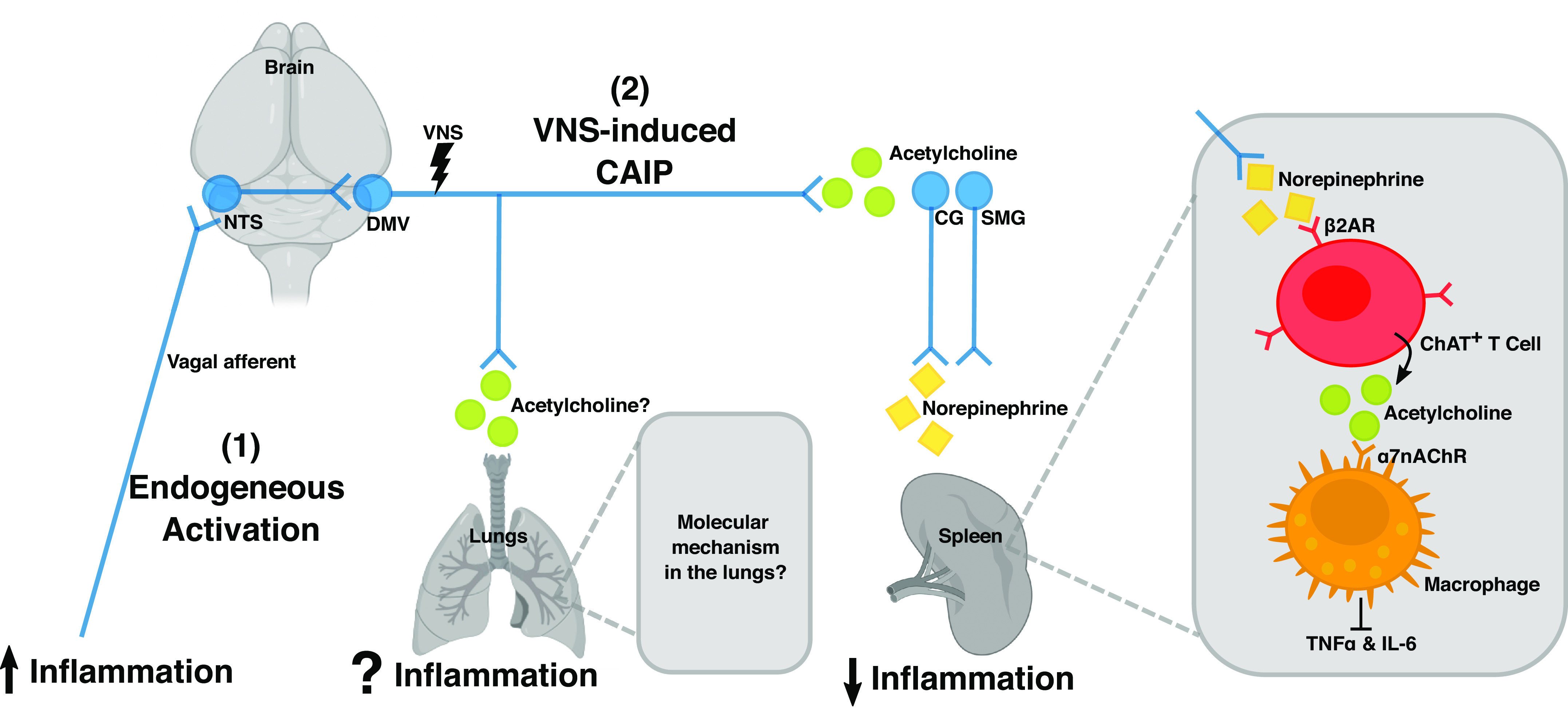
Figure 1.

A schematic illustration of the cholinergic anti-inflammatory pathway (CAIP) in the spleen. Vagal efferent fibers activate sympathetic neurons in the superior mesenteric ganglia (SMG) or celiac ganglia (CG) that project into the spleen. This leads to a local release of norepinephrine, which induces choline acetyltransferase (ChAT)-positive T cells to release acetylcholine. Acetylcholine in turn activates macrophages by binding to the nicotinic α 7 receptor (α7nChR), which inhibits the production of proinflammatory cytokine, such as TNFα and IL-6. Together, these cells constitute the CAIP, and selective efferent VNS modulates inflammation in the spleen via this pathway. It remains elusive if the same neuroimmune circuit is present in the lungs. [Image created with BioRender.com and published with permission.]
