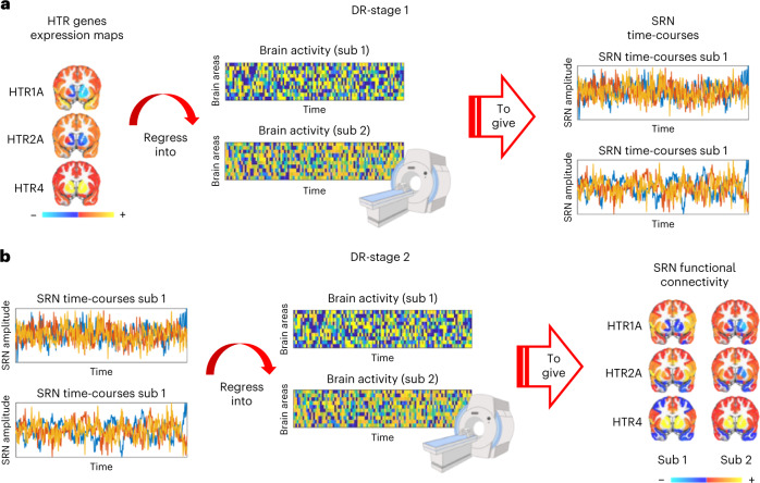
Fig. 1. Mapping functional signatures of brain-wide SRNs.
Gene expression brain maps of serotonin receptor genes are combined with fMRI (both mouse and human) via FSL DR to map temporal and spatial fMRI signatures of different serotonin receptor types. a, DR-stage 1: regresses the spatial maps of serotonin receptor genes into each subject’s four-dimensional (4D) fMRI dataset. This gives a subject-specific time-course quantifying network amplitude changes for each SRN. DR-stage 1 can be used to address questions such as: What are the SRN amplitude changes in response to optogenetic stimulation? b, DR-stage 2: regresses subject-specific time-courses into the same 4D fMRI dataset. This provides a subject-specific spatial map quantifying functional connectivity for each SRN. DR-stage 2 can be used to address questions such as: What are the SRN brain correlates associated with individual differences in a depression scale? Sub, subject.

