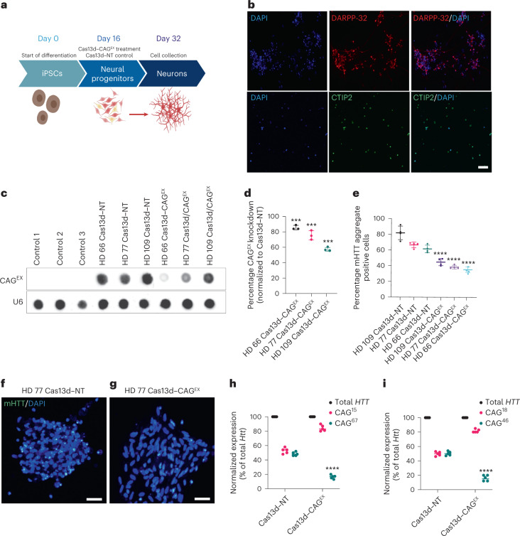
Fig. 2. Cas13d–CAGEX reduces mHTT mRNA and protein in cells derived from patients with HD.
a, Schematic of the differentiation protocol used for iPSC-derived neurons treated with a lentiviral system expressing Cas13d–CAGEX. b, Representative immunofluorescence images of cells at day 32 in control neuronal cells, showing that the cells are positive for striatal markers, DARPP-32 (top) and CTIP2 (bottom). Scale bar, 20 μm. n = 1 technical replicate. c,d, RNA dot blot analysis (c) and quantification (d) of CAG-expanded RNA within MSN cells transduced with Cas13d–NT- or Cas13d–CAGEX-expressing lentiviral vectors (one-way ANOVA, Tukey post hoc test, ***P < 0.001). HD 66 Cas13d–CAGEX P = 0.00037, HD 77 Cas13d–CAGEX P = 0.00054, HD 109 Cas13d–CAGEX P = 0.00067; n = 3 technical replicates, n = 3 biological replicates. Data are presented as mean values ± s.e.m. e–g, Quantification of mHTT aggregates within MSN cells transduced with Cas13d–NT- or Cas13d–CAGEX-expressing lentiviral vectors (e) (one-way ANOVA, Tukey post hoc test, ****P < 0.0001, n = 3 technical replicates, n = 4 differentiations, 200 cells per experimental group). Representative images of the analysis are in f and g (scale bar, 20 μm). Data are presented as mean values ± s.e.m. h,i, Quantification of mutant and WT HTT RNA via allele-specific RT-qPCR in GM04723 (h) and GM02151 (i) fibroblasts (two-way ANOVA, Tukey post hoc test, ****P < 0.0001, n = 5 biological replicates). DAPI, 4,6-diamidino-2-phenylindole.

