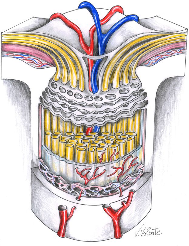
Fig. 1. The optic nerve at the lamina cribrosa level.

Artistic drawing by Vinicio Valente, neurosurgeon at the “Annunziata” Hospital, Cosenza, Italy, illustrating a section of the optic nerve at the lamina cribrosa level, which forms the anatomical floor of the optic nerve head and separates the intraocular and intracranial pressure compartments. Lowering of intracranial pressure, by cerebrospinal fluid shunting in idiopathic normal pressure hydrocephalus may increase the pressure gradient across the lamina cribrosa leading to glaucomatous damage.
