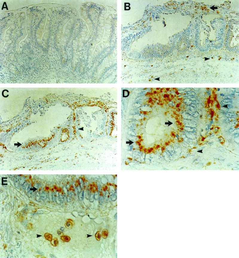
FIG. 2.
Immunohistochemistry for Cox-2 (A, C, D, and E) and SREHP (B and E). (A) Cox-2 immunohistochemistry in uninfected xenograft. (B) Immunohistochemistry for SREHP 24 h after infection with E. histolytica trophozoites. Trophozoites are in debris over an ulcer (arrow) and in the submucosa (arrowheads). (C) Cox-2 immunohistochemistry in a xenograft 24 h after infection with trophozoites. There is staining of crypt epithelial cells (arrow) and of cells in the lamina propria (arrowhead). (D) High-power view of an area in panel C showing Cox-2 staining in crypt epithelial cells (arrow) and lamina propria cells (arrowheads). (E) Staining for Cox-2 in crypt epithelial cells (arrow) and staining for SREHP (arrowheads). Magnification, ×200 (A through C) and ×1,000 (D and E).

