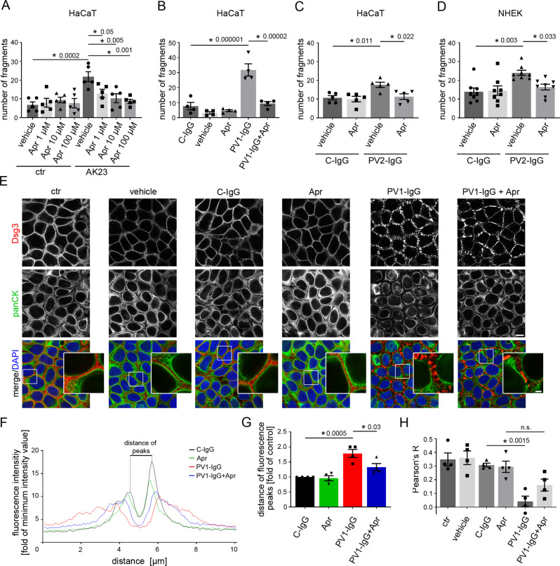
Fig. 2. Apremilast ameliorates PV-IgG-induced loss of intercellular adhesion and keratin filament alterations.
A–D Dispase-based keratinocyte dissociation assay of human keratinocytes. 1 h pre-incubation of apremilast attenuated anti-Dsg3 antibody (AK23) in a dose-dependent fashion (A, n = 5). 100 µM apremilast also ameliorated PV1-IgG- (B, n = 4) and PV2-IgG- (C, n = 5) induced loss of cell cohesion in HaCaT cells. D Primary keratinocytes (NHEK) show same results with PV2-IgG. (n = 8). E Immunostaining of Dsg3 and keratin filaments (panCK) in HaCaTs. Apremilast (100 µM) ameliorated PV1-induced keratin retraction but not Dsg3 depletion and fragmentation of staining. Scale bar = 10 µm. Zoom in areas marked with white rectangles. Scale bar (zoom) = 2.5 µm. Representative of n = 4. F, G Quantification of cytokeratin fluorescence intensity in small areas perpendicular to the respective cell border. F Average of keratin fluorescence intensity measured along 10 µm spanning a cell border under respective conditions. G Apremilast improved PV1-IgG-induced increase in distance of fluorescence peaks as a measure for retraction of the keratin cytoskeleton (n = 4). H Calculation of Pearson’s correlation coefficient. Apremilast ameliorated PV1-IgG induced loss of co-localization between keratin filaments and Dsg3 (n = 4). Columns indicate mean value ± SEM, *P < 0.05 (exact values are depicted in the figure). One-way ANOVA with Bonferroni correction. Source data are provided as a Source Data file.

