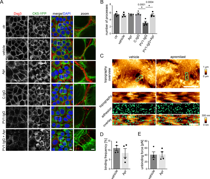
Fig. 3. Apremilast restores PV1-IgG-induced retraction of the keratin cytoskeleton but has no effect on Dsg3.
A Immunostaining of Dsg3 in stably expressing CK5-YFP HaCaTs. Apremilast (100 µM) prevented PV-IgG-induced retraction of the keratin cytoskeleton but not Dsg3 depletion and fragmentation of Dsg3 staining. Representative of n = 4. Scale bar = 10 µm. Areas for zoom in marked with white rectangles. Scale bar (zoom) = 2.5 µm. B Quantification of the number of keratin-bearing processes over 10 µm of the membrane. Pretreatment of apremilast restored PV1-IgG-induced decreased number of keratin-bearing cell processes (n = 4). C Topography overview images of atomic force microscopy (AFM) measurements on living HaCaTs using a Dsg3-functionalized tip revealing cell borders bridged by dense filamental structures. Scale bar = 10 µm. Small areas along the cell borders (green rectangles) were chosen for adhesion measurements. In these areas each pixel represents a force-distance-curve. In the adhesion panel each green pixel represents a Dsg3-specific binding event. Scale bar = 1 µm. D, E Quantification of AFM adhesion measurements. D Apremilast (100 µM) had no effect on Dsg3 binding frequency. Additionally, the unbinding force (E) as a measure for the single molecule binding strength was unaltered in cells treated with apremilast (8 cell borders from 4 independent experiments with 900 force-distance curves/ cell border). Bars indicate mean value ±SEM. *P < 0.05 (exact values are depicted in the figure). One-way ANOVA with Bonferroni correction (B), two-tailed Student’s t test (D, E). Source data are provided as a Source Data file.

