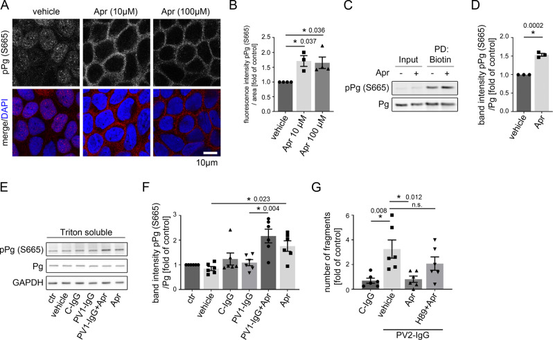
Fig. 4. Apremilast increases phosphorylation of Pg at S665.
A Immunostaining of phosphorylated Pg at S665 in HaCaT showed increased phosphorylation of Pg at S665 upon apremilast (10 and 100 µM) treatment. Representative of n = 4. Scale bar = 10 µm. B Quantification confirms significant stronger pPG (S665) staining along keratinocyte cell borders (n = 4). Bars indicate mean value ±SEM. C Biotinylation assay reveal higher levels of pPg (S665) at the cell membrane of HaCaT keratinocytes after apremilast (10 µM) compared to control. Representative of n = 3. D Quantification of the biotin pulldown of the biotinylation assay. Representative Western blot of HaCaT Triton soluble fraction (E) and quantification of band intensity of pPg (S5665) (F) show a significant increased phosphorylation of Pg at S665 after apremilast (100 µM) treatment. GAPDH and total Pg was used as loading control (n = 6). G Inhibition of PKA (H89) inhibited the protective effect of apremilast (10 µM) on PV2-IgG-induced loss of cell adhesion in keratinocyte dissociation assay in HaCaTs (n = 6). Columns indicate mean value ±SEM. *P < 0.05 (exact values are depicted in the figure). Two-tailed Student’s t test (D), One-way ANOVA with Bonferroni correction (B, F, G). Source data are provided as a Source Data file.

