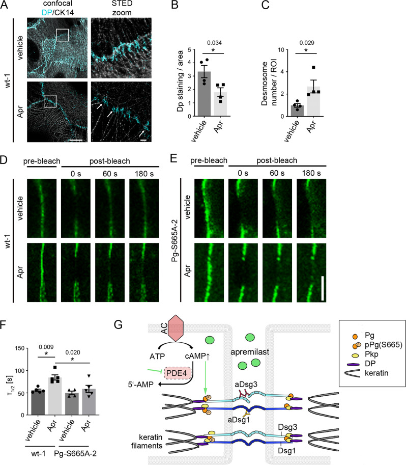
Fig. 7. Apremilast regulates desmoplakin recruitment and Dsg3 membrane availability.
A Representative immunostaining of wt murine keratinocytes incubated with apremilast for 2 h and stained for desmoplakin (Dp) and cytokeratin 14 (CK14). Under control conditions, Dp was present at linear streaks along the cytokeratin filament at cellular contact points. In contrast, after apremilast (10 µM) treatment, Dp was present in desmosomes (white arrows). Scale bar = 10 µm. Zoom in areas marked with white rectangles. Scale bar (zoom) = 1 µm. Representative of n = 4. B, C Quantification of Dp staining. Dp-covered area is significantly reduced after apremilast treatment compared to control cells. In contrast, number of desmosomes/ROI was elevated. D, E FRAP on murine keratinocytes transfected with Dsg3-GFP. D, F In wt murine keratinocytes, apremilast (10 µM) lead to enhanced recovery halftime (τ1/2). Scale bar = 5 µm. E, F In contrast, no changes in τ were present in Pg-S665A keratinocytes (n = 5, for Pg-S665A vehicle: n = 4). G Schematic of mechanisms of protective cAMP signaling in pemphigus. Apremilast prevents PV-IgG-induced loss of intercellular adhesion. Autoantibodies directed against desmogleins induce uncoupling of keratin filaments from the desmosomal plaque. Increase of intercellular cAMP by the phosphodiesterase 4- (PDE4) inhibitor apremilast ameliorates this effect by PKA-dependent phosphorylation of Pg at S665. Columns indicate mean values ±SEM. *P < 0.05 (exact values are depicted in the figure). Two-tailed Student’s t test (B, C), one-way ANOVA with Bonferroni correction (F). Source data are provided as a Source Data file.

